Neurotechnology
Electroencephalography (EEG): Signals, Uses and Configurations
Electroencephalography records voltage differences at the scalp that reflect the summed electrical activity of large neuronal populations. Its defining advantage is temporal resolution: EEG can follow brain dynamics on the scale of milliseconds, which makes it central to clinical neurophysiology, cognitive neuroscience, neurofeedback and many brain–computer interfaces.
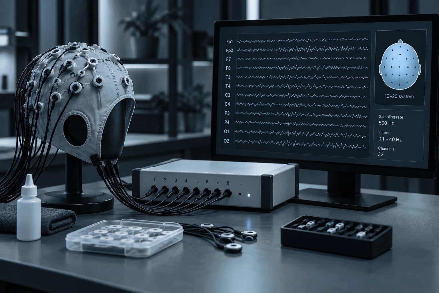
The physiological basis of the EEG signal
Scalp EEG measures electrical potential differences generated largely by synchronized postsynaptic activity in neuronal populations. These signals reach the electrodes after propagating through brain tissue, cerebrospinal fluid, skull and scalp, so the recorded activity reflects population-level electrophysiology and is spatially shaped by volume conduction, the spread of electrical fields through brain tissue, cerebrospinal fluid, skull and scalp.
EEG systems commonly analyze spontaneous rhythms, event-related potentials (ERPs), brief voltage changes time-locked to a stimulus or event and changes in spectral power, which describes how strongly EEG activity is represented within a defined frequency band or connectivity. Interpretation depends on montage, reference, electrode quality, filtering, sampling and the experimental or clinical context. [1–3]
Clinical EEG and research EEG have different goals
Routine and sleep EEG follow formal recording standards because their purpose can include clinical interpretation, especially in epilepsy and related neurophysiology. Long-term video-EEG adds synchronized behaviour and video to electrophysiology. [1,4]
Research EEG may instead prioritize high-density coverage, mobile recording, precise stimulus timing, source analysis or integration with VR, eye tracking, EMG or stimulation. The value of a higher channel count depends on the scientific question, spatial coverage and planned analyses.
Typical EEG use cases
EEG is used when the timing of brain activity matters. The same basic measurement can support very different applications depending on montage, recording duration, task design and analysis.
- Routine, sleep and long-term video EEG
- Event-related potentials and cognitive experiments
- Motor-imagery, P300 and SSVEP BCI
- EEG neurofeedback
- Mobile, VR and movement studies
Why EEG is important for BCI
EEG is the dominant non-invasive BCI signal because it is comparatively accessible, portable and fast. For example, motor-imagery BCIs use changes in sensorimotor rhythms—EEG rhythms over sensorimotor cortex that change during real or imagined movement. P300-based BCIs use a positive EEG response that often becomes prominent roughly 300 milliseconds after a task-relevant or infrequent stimulus. Steady-state visual evoked potential (SSVEP) BCIs use rhythmic EEG responses that follow the frequency of a periodically flickering visual stimulus. [5]
The challenge is that EEG is sensitive to eye movements, facial and neck muscle activity, cable movement, poor electrode contact and environmental electrical noise. Good BCI performance therefore depends as much on acquisition quality and experimental design as on the classifier.
Common EEG configurations
EEG systems range from compact few-channel mobile setups to 32- or 64-channel general research systems and 128-channel or higher-density arrays. Configurations also differ in electrode technology, mobility and access to real-time data.
- Wet, semi-dry or dry electrodes
- Active or passive electrodes
- Wired or wireless/mobile amplifiers
- Compact, 32/64-channel and high-density systems
- Trigger inputs, raw-data access and real-time streaming
Evidence
References and further reading
- 1.Peltola ME et al. Routine and sleep EEG: Minimum recording standards of the IFCN and ILAE. Clinical Neurophysiology, 2023.
- 2.Nuwer MR et al. IFCN standards for digital recording of clinical EEG. Electroencephalography and Clinical Neurophysiology, 1998.
- 3.Babiloni C et al. IFCN EEG research workgroup recommendations on frequency and topographic analysis of resting-state EEG rhythms. Clinical Neurophysiology, 2020.
- 4.Tatum WO et al. Minimum standards for inpatient long-term video-EEG monitoring. Clinical Neurophysiology, 2022.
- 5.Wolpaw JR et al. Brain-computer interfaces for communication and control. Clinical Neurophysiology, 2002.
Next step
Interested in this technology?
Contact me to discuss available options and suitable configurations.
Contact me about this technologyContinue
Related articles
Neurotechnology
Brain–Computer Interfaces (BCI)
A brain–computer interface measures central nervous system activity and translates it into an artificial output that can communicate, control a device, or provide feedback without relying entirely on the usual peripheral motor pathway. Modern BCIs range from communication syst…
Neurotechnology
Functional Near-Infrared Spectroscopy (fNIRS)
Functional near-infrared spectroscopy is a non-invasive optical method that estimates relative changes in oxygenated and deoxygenated haemoglobin using near-infrared light. Portable systems make it possible to study cortical haemodynamics in settings where MRI is impractical o…
Neurotechnology
Experimental Response and Synchronization Systems
Neuroscience experiments need more than biological recording: they also need an accurate account of when each stimulus appeared, when the participant responded, and how those events align with signals collected by different devices. Response hardware, event markers and synchro…