Neurotechnology
Functional Near-Infrared Spectroscopy (fNIRS): Uses and Configurations
Functional near-infrared spectroscopy is a non-invasive optical method that estimates relative changes in oxygenated and deoxygenated haemoglobin using near-infrared light. Portable systems make it possible to study cortical haemodynamics in settings where MRI is impractical or too restrictive.
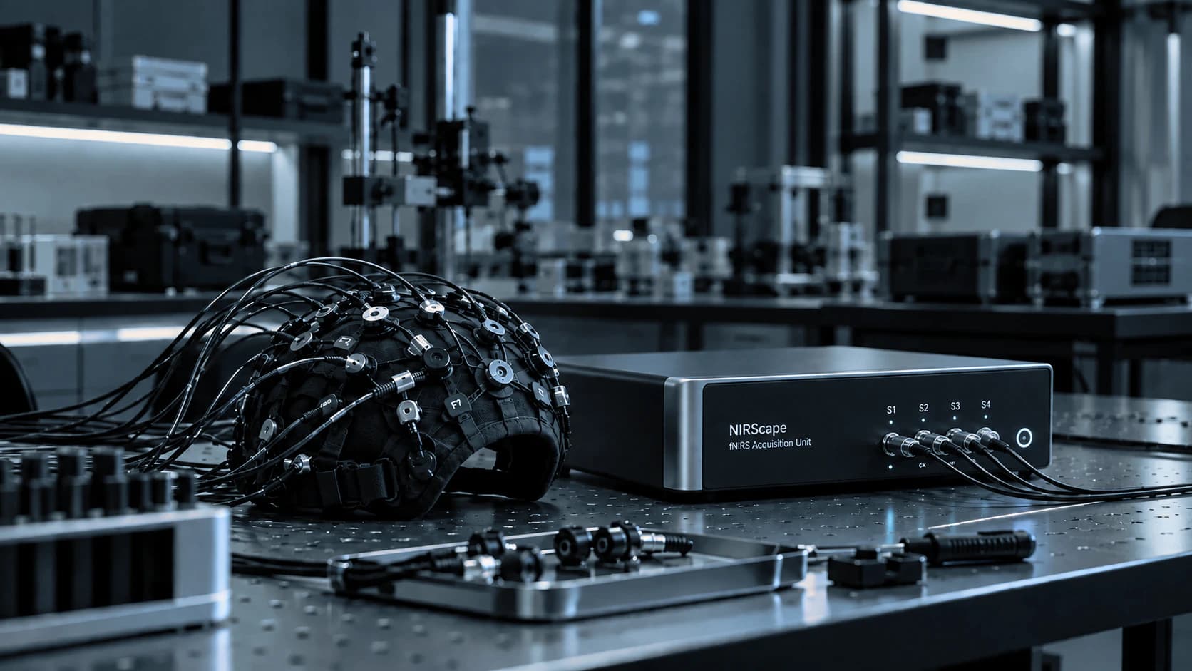
Physical and physiological basis of the fNIRS signal
Near-infrared light can pass through scalp and skull tissue, while oxygenated and deoxygenated haemoglobin absorb the light differently. Source–detector pairs placed on the head are used to estimate haemodynamic changes in superficial cortical regions. [1,2]
The fNIRS signal is linked to neurovascular coupling—the process by which local neural activity is followed by changes in blood flow and oxygenation—and therefore reflects vascular responses associated with neural activity rather than direct electrophysiological activity. The haemodynamic response develops over seconds, so its temporal resolution is substantially slower than EEG.
Mobility and naturalistic research
Compared with functional MRI, fNIRS can be used in more mobile and natural environments. It has been applied to social interaction, walking, rehabilitation tasks, infant development and other naturalistic research settings. [2,3]
Hyperscanning means recording brain activity from two or more people at the same time while they interact or perform a shared task. It allows researchers to quantify inter-brain coupling metrics, but interpretation requires careful controls for common task structure, chance correlations and systemic physiology. [6]
Typical fNIRS use cases
fNIRS is especially useful when cortical haemodynamics need to be measured outside the constraints of an MRI scanner or during interaction and movement.
- Cognitive and developmental neuroscience
- Walking and mobile-task studies
- Social interaction and hyperscanning
- Rehabilitation and motor tasks
- Simultaneous EEG–fNIRS research
Systemic physiology and extracerebral contributions
Changes in blood pressure, respiration, heart rate and superficial scalp blood flow can contaminate fNIRS signals. Poor optode contact, hair and motion can create additional artefacts. Best-practice guidance therefore emphasizes optode geometry, signal quality, preprocessing, reporting and methods such as short-separation channels, in which source–detector pairs are placed close together to capture mainly superficial scalp physiology or concurrent systemic physiology. [3–5]
This is especially important when claims concern subtle cognitive effects. Accordingly, haemoglobin changes require analysis that separates cortical responses from systemic and extracerebral contributions.
fNIRS with EEG and BCI
EEG and fNIRS are complementary: EEG captures fast electrical dynamics; fNIRS captures slower haemodynamic dynamics. Simultaneous systems can be valuable when a study needs both, and hybrid EEG–fNIRS BCIs are an active area of development. [7]
The trade-off is complexity. Headgear layout, timing, shared triggers, motion artefacts and analysis pipelines must be designed as one system rather than as two independent devices.
Common fNIRS configurations
fNIRS systems differ in optical architecture, number and arrangement of sources and detectors, portability and whether short-separation measurements or multimodal integration are available.
- Regional sparse-channel measurements
- High-density optical arrays
- Wearable or wireless caps
- Short-separation channels for superficial physiology
- Hyperscanning or simultaneous EEG–fNIRS setups
Evidence
References and further reading
- 1.Ferrari M, Quaresima V. A brief review on the history of human functional near-infrared spectroscopy development and fields of application. NeuroImage, 2012.
- 2.Pinti P et al. The present and future use of functional near-infrared spectroscopy for cognitive neuroscience. Annals of the New York Academy of Sciences, 2020.
- 3.Yücel MA et al. Best practices for fNIRS publications. Neurophotonics, 2021.
- 4.Tachtsidis I, Scholkmann F. False positives and false negatives in functional near-infrared spectroscopy. Neurophotonics, 2016.
- 5.Scholkmann F et al. Systemic physiology augmented functional near-infrared spectroscopy. Neurophotonics, 2022.
- 6.Quantification of inter-brain coupling: a review of methods used in haemodynamic and electrophysiological hyperscanning studies, 2023.
- 7.Simultaneous functional near-infrared spectroscopy and electroencephalography for monitoring human brain activity and oxygenation: a review, 2017.
Next step
Interested in this technology?
Contact me to discuss available options and suitable configurations.
Contact me about this technologyContinue
Related articles
Neurotechnology
Electroencephalography (EEG)
Electroencephalography records voltage differences at the scalp that reflect the summed electrical activity of large neuronal populations. Its defining advantage is temporal resolution: EEG can follow brain dynamics on the scale of milliseconds, which makes it central to clini…
Neurotechnology
Brain–Computer Interfaces (BCI)
A brain–computer interface measures central nervous system activity and translates it into an artificial output that can communicate, control a device, or provide feedback without relying entirely on the usual peripheral motor pathway. Modern BCIs range from communication syst…
Neurotechnology
Experimental Response and Synchronization Systems
Neuroscience experiments need more than biological recording: they also need an accurate account of when each stimulus appeared, when the participant responded, and how those events align with signals collected by different devices. Response hardware, event markers and synchro…